Kitware customer highlight: SonoVol
At Kitware, we provide collaborative research, development, and technology integration services for companies that work in the medical and biomedical business sectors. We work with such companies to streamline their internal software and data analysis processes, generate prototypes to raise venture capital or conduct first-in-human trials, and investigate algorithms to upgrade existing commercial offerings.
As part of a series that highlights many of the applications that we developed in collaboration with our customers, this post features our joint research and development efforts with SonoVol. These efforts focused on building a prototype of a high-throughput, ultrasound-based, optical imaging system for small animals to accelerate preclinical drug research.

SonoVol: The Company
SonoVol is a spin-off company from the University of North Carolina at Chapel Hill. The company is building an automated, high-throughput, ultrasound-based, small-animal imaging system to accelerate the pace and reduce the cost of research in preclinical disease and drug development. This desktop system will allow researchers to generate quantitative metrics of treatment efficacy from longitudinal structural and functional imaging data. The imaging system will be a powerful tool for cancer and cardiovascular research projects in small and large university laboratories, government laboratories, and pharmaceutical companies.
The SonoVol imaging system combines multiple advanced hardware and software components. The company’s work with Kitware minimized the complexity of the user interface of the system for high-throughput, longitudinal studies. The work maximized the accuracy and stability of the measures the system produces. The graphical user interface must intuitively coordinate acquisitions from ultrasound transducers and optical devices, precisely control motors to create consistent whole-body imagery, and maintain calibration via tracking sensors and customized hardware. Advanced quantification and visualization software algorithms then fuse and analyze the data.
Need: Why SonoVol came to Kitware
SonoVol partnered with us to build upon our expertise in image analysis and software engineering. The prototype is built on our open-source platforms 3D Slicer, the Insight Segmentation and Registration Toolkit (ITK), and the Visualization Toolkit (VTK). 3D Slicer provides a foundation for the management and visualization of three-dimensional (3D) medical images from multiple modalities. ITK provides the image quantification methods, such as the abilities to reconstruct ultrasound data (ITK Ultrasound), register multiple ultrasound sweeps to form a whole-body image, and segment anatomic structures from the data. VTK provides multi-modality visualization capabilities as well as interactive widgets to specify regions of interest, place annotations, and graph results.
Our work was highly collaborative; both groups were committed to the success of the project. We held frequent design meetings with the SonoVol team. Our joint focus was on testing to determine system precision, system calibration drift, and potential imaging protocols. We maintained careful animal welfare considerations. During implementation, we also engaged with hardware manufacturers to address issues that we discovered with device drivers and application programming interfaces (APIs). As a strong team, we shifted task priorities and effort allocations seamlessly to meet evolving demands for demonstrations and to address the many challenges that came up during the development of this innovative product.
The collaborative innovation that permeated the rapid development of the SonoVol product exemplifies many of the strengths of Kitware. By building on 3D Slicer, ITK, and VTK, this product could go from concept to implementation in months instead of years. The final prototype has demonstrated a level of precision and stability that is typically found only in well-matured products. While clearly driven by the management and engineers of SonoVol, these successes can also be attributed to our domain knowledge in image analysis, ultrasound, tracking, volumetric image formation, multi-modality registration, image quantification, and biomedical research. In addition, this project benefited from our expertise in software practices, such as test-driven development, nightly regression tests, and design patterns and processes for scalable and maintainable software systems. Furthermore, we provided expertise in other aspects of commercial application development, such as customized user interfaces, interactive visualizations, high-performance computing, and software packaging.
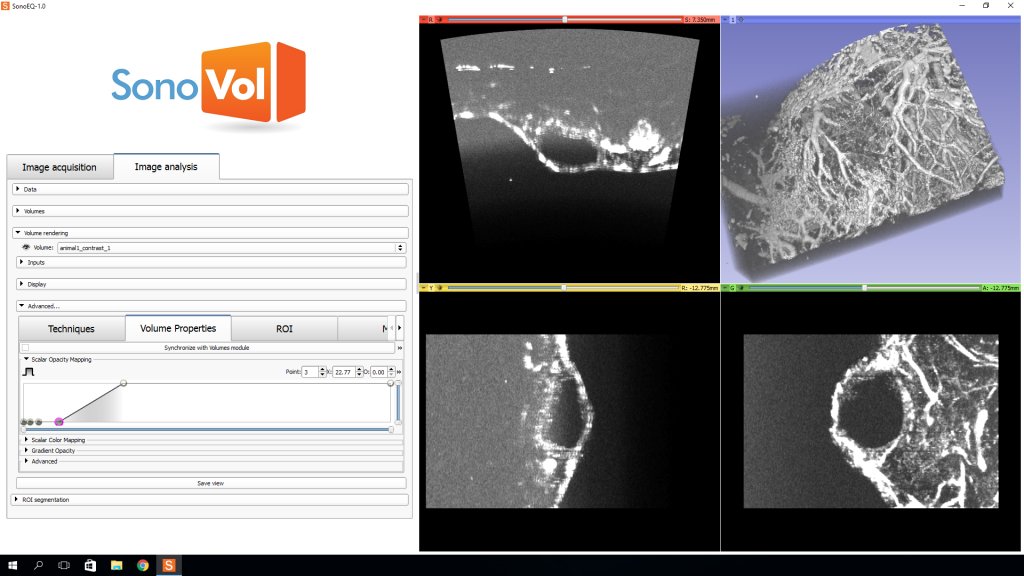
Custom application developed by Kitware
As shown in the above figure, we helped SonoVol develop the software for their preclinical imaging system. We used 3D Slicer to rapidly prototype the SonoVol application to control and analyze image and sensor data, provide an end-user graphical application that is capable of 3D visualization, and communicate and integrate all of the software components. The application is currently under development, and preclinical validation studies are anticipated to begin soon.
To learn more about how we can tailor solutions to meet your research needs, please contact kitware@kitware.com.