Emerging Trends in Digital Dentistry
A lot of domains in healthcare are rapidly shifting from manual, experience-driven workflows to hybrid, AI-guided, human in the loop workflows. Advances in imaging, sensors, and AI are allowing measurement, diagnosis and documentation tasks to happen faster than ever before.
There is a large body of research and a wide array of available open source tools looking to automate several diagnostic tasks in clinical dentistry. The field has evolved thanks to the availability of new data acquisition techniques, the ability to merge multimodal datasets, the globalization and democratization of dental datasets and, of course, the prevailing presence of AI. We wanted to take a look at the landscape in digital dentistry and discuss the different research patterns that emerge that can help us understand where the field can go next.
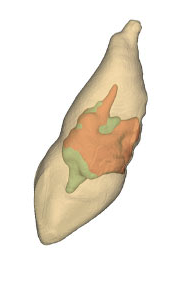
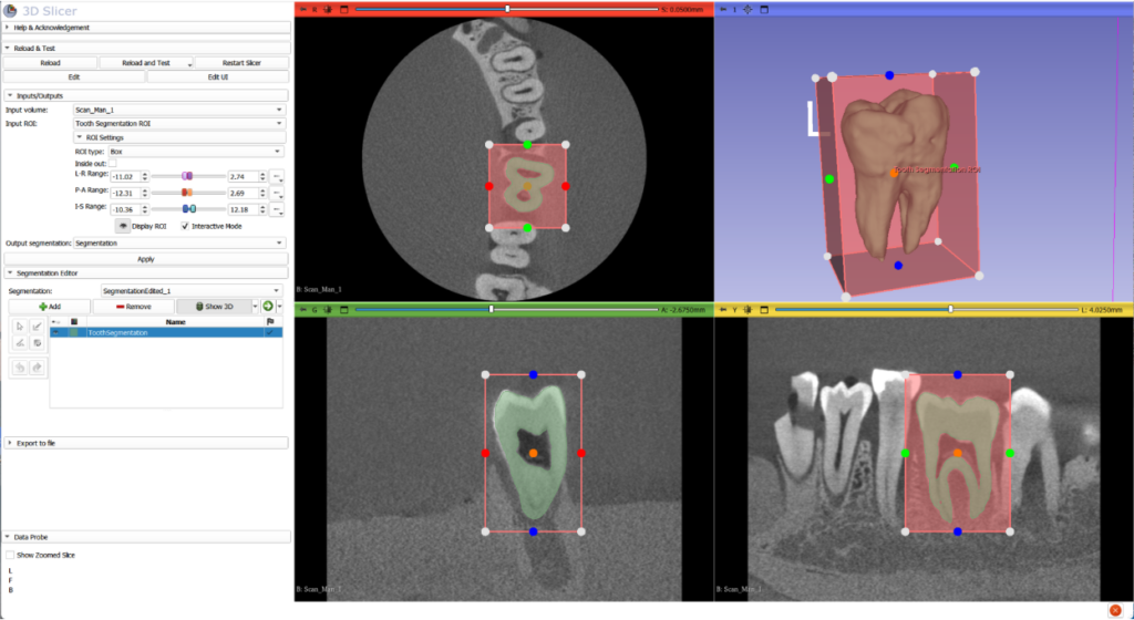
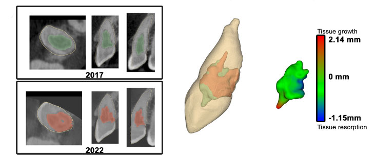
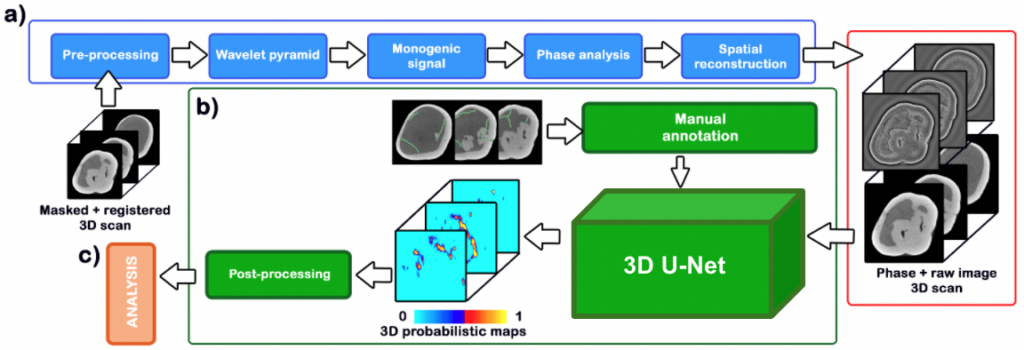
Tooth Detection: Automatic tooth detection continues to be an important focus in digital dentistry. The goal of this task is to isolate teeth in different image acquisition modalities both for internal analysis as well as for understanding their relationship with the surrounding dentition and the rest of the skeleton. Thanks to advances in AI-based image segmentation, there is a large amount of tooling available, both for research and commercial purposes, that allows to accurately isolate and locate teeth in different types of images. Tooth isolation can be challenging in the presence of noisy data that includes acquisition artifacts, low resolution scanners and presence of metal implants. Once the tooth is segmented, it is possible to analyze its internal structure to detect diseases of the dentin and pulp, tissue loss or fractures. It is also possible to study its structure and how it relates to other teeth as well as the maxilla and mandible.
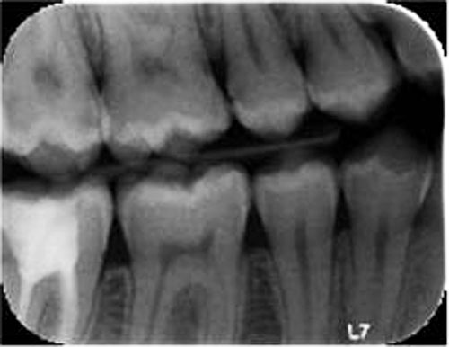
Inferring 3D from 2D: Even when it is clear that understanding dental health in three dimensions is the goal of any dental diagnostic task, bitewing radiograph is still the standard of care to diagnose dental pathologies because of its lowered radiation exposure, wide availability, speed and low cost. Thanks to advances in shape analysis, AI and more widely available 3D datasets, it is possible to derive 3D information from inherently 2D imaging techniques. Our team has performed extensive work in this area, see more information here.
Soft tissue analysis: Beyond tooth health there are a number of serious oral health problems that can affect the oral mucosa as well as the tongue. The field of dermatology has spearheaded the use of natural light photography for the detection of skin lesions, and there is real potential in using it in oral care for the detection of mucosal lesions, tongue abnormalities, oral cancers or gingival inflammation and recession.
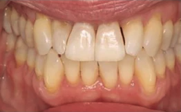
Connected personal devices: Clinicians and researchers in dentistry have started to use natural-light or smartphone photography in the detection and monitoring of mucosal lesions, tongue abnormalities, and oral cancers. In clinical practice, the images are still only used for documentation and referral. In research, AI models trained on these images are demonstrating promising performance for automated screening, particularly in low-resource or telehealth contexts.
All of these incipient technologies are being powered by dental images and AI, but there is much more to digital dentistry than just images. By using just images, AI-identified findings will stay within the structural domain, when other important clinical connections can exist. AI models that can combine data across modalities hold enormous potential to improve feature representation within those data domains and unlock new discoveries. Electronic Health Records, diagnostic notes and clinical systemic variables all can be incorporated to existing structural AI models that can predict treatment responses and other outcomes. Data integrity checks for deep learning datasets, independent of modality, will ensure dataset accuracy, consistency, and reliability, preventing issues like model drift, bias, and poor performance. Essential checks include validating for missing values, duplicates, label accuracy, and schema consistency, alongside with grounding strategies as well as monitoring training-test distribution shifts. These steps will ensure data remains fit for training high-quality, trustworthy models.
Dental Software at Kitware
Kitware’s team has years of experience in Digital Dentistry. Our team has performed multimodal reconstruction, implant design, pathology/disease monitoring, surgical planning and simulation, and developed applications alongside our commercial partners that are currently being used in clinical practice.
In research, we have contributed to a number of open source toolkits in the field such as SlicerCMF, or DentalSegmentator. These toolkits have assisted researchers in generating groundbreaking findings in Digital Dentistry.

How Kitware Can Help
Digital Dentistry is evolving from image-based expert diagnosis into a connected, semi-automated multimodal, and clinically integrated field. What began with tooth detection and segmentation has expanded into 3D anatomical reasoning, soft-tissue screening through photography, and AI systems that support monitoring beyond the clinic walls. Advances in AI combined with 3D anatomical reconstruction, screening through photography and data fusion will move the field closer to true decision support rather than isolated unimodal analysis.
Kitware is at the front end of research that includes high-volume, high-quality data, human oversight and seamless integration with real clinical workflows. We know that the future of digital dentistry lies in AI systems that enhance clinical expertise, with the goal of delivering more precise diagnostics, earlier detection, and more personalized care for patients worldwide.
Contact us to learn how we can help you design your automated workflows and integrate our open-source platforms and libraries into your Digital Dentistry solutions for research and products.