Delivering Innovation Through Custom Medical Imaging Software
RSNA is the largest radiology conference in the U.S. featuring the latest in CT, MRI, AI, 3D printing and other imaging modalities. As an industry leader in using AI and machine learning to analyze medical images and data, Kitware is exhibiting at RSNA 2022 to showcase our latest radiology platforms.
We look forward to meeting with attendees and other exhibitors during the conference. If you are attending, be sure to visit our booth #4354 to see demos of our state-of-the-art medical image analysis solutions.
Learn About Our Latest Open Source Radiology Platforms
Schedule an introductory meeting to see our state-of-the-art medical image analysis solutions and how we can work together to apply them to your research or products.
Kitware’s Medical Computing Open Source Platforms


Learn more about our custom medical computing software
Artificial Intelligence Showcase
Artificial Intelligence Models for prediction of Bronchopulmonary Dysplasia in Neonates
Link opens in a Google Slide. Slides will rotate every 30 seconds or users can use the mouse to click forward. Left and Right arrow keys also navigate through the slides.
Dental Fracture Detection
Link opens in a Google Slide. Slides will rotate every 30 seconds or users can use the mouse to click forward. Left and Right arrow keys also navigate through the slides.
Medical Image Quality Assurance using Deep Learning
Link opens in a Google Slide. Slides will rotate every 30 seconds or users can use the mouse to click forward. Left and Right arrow keys also navigate through the slides.
Open Science for the Challenges of Medical Imaging AI
Link opens in a Google Slide. Slides will rotate every 30 seconds or users can use the mouse to click forward. Left and Right arrow keys also navigate through the slides.
Point of care Ultrasound
Link opens in a Google Slide. Slides will rotate every 30 seconds or users can use the mouse to click forward. Left and Right arrow keys also navigate through the slides.
Kitware’s Medical Computing Areas of Expertise
3D Slicer-Based Applications
We create custom plugins, SDKs, applications, and software packages using 3D Slicer. 3D Slicer has been used in a variety of medical and basic scientific applications such as dentistry, radiation oncology, surgical planning, and drug development. These custom software applications can be deployed to local hardware or to remote servers using Docker or on tablets. To support reproducible workflows, they can also be integrated with our Girder data management solution or with Jupyter notebooks.
Bioinformatics
Our solutions connect cutting-edge bioinformatics research to a solid, extensible platform, where open science is front and center. We develop data management, analysis management, and visualization applications for a wide range of areas including genomics, metabolomics, and phylogenetics. From browsing and analyzing large histology slides on the web, to stabilizing and sharing new ‘omics algorithms through a robust web application, we have data and analytics covered.
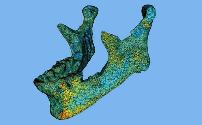
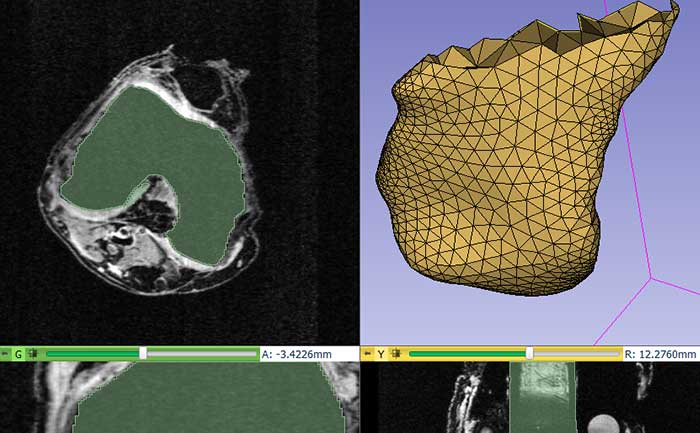
Dental, Craniomaxillofacial, and Musculoskeletal Image Analysis
Our projects and research aim to quantitatively explore how age, disease or treatment affect structures in the skeleton and craniomaxillofacial (CMF) complex. This improved knowledge can help diagnose disease early, plan and measure treatment, and monitor the progression of certain conditions. In particular, we are experts in morphometry analysis, a technique that can be used to quantitatively plan surgery or measure remodeling in the bones. Our musculoskeletal image analysis methods, for example, can quantify bone quality or tooth integrity. We also develop CMF-specific surgical trainers to improve procedural knowledge and surgical proficiency without sacrificing patient safety.
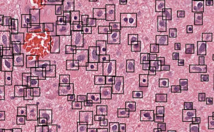
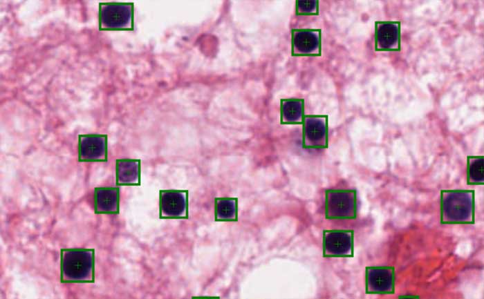
Digital Pathology
We are building a suite of open source web-based informatics tools that manage, visualize, and analyze massive and growing collections of data in digital pathology. The key solutions in the making include Digital Slide Archive (DSA), HistomicsTK, and Large-image. DSA is a web-based platform for the aggregation, management, and dissemination of large collections of whole-slide histopathology images, along with associated clinical and genomic metadata. HistomicsTK serves as both a web-based analytics platform and a standalone Python toolkit. It contains computer vision and machine learning algorithms for the quantitative analysis of whole-slide histopathology images and associated data. Large-image supports the web-based visualization and annotation of large multi-resolution whole-slide histopathology images. It also includes a Python API for reading/writing these images in a tiled fashion.
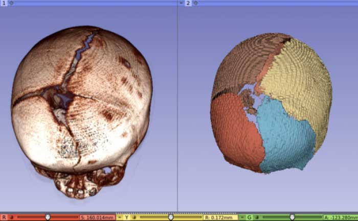
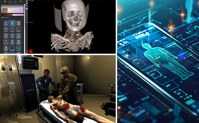
Image Guided Intervention and Surgical Planning
We develop image-guided intervention and surgical planning applications that replace traditional surgery and invasive procedures with minimally invasive techniques that incorporate medical imaging to guide the intervention. Patients prefer these procedures to open surgeries because they are typically less traumatic to the body and result in faster recovery times. Technological advancements in medical imaging, registration algorithms, visualization technologies, and tracking systems are driving forces behind increased adoption of these procedures by physicians. Software is an integral part of these image-guided intervention systems. Whether it is for interfacing with a tracking device to collect position information from surgical instruments, integrating intraoperative and pre operative images, or generating a 3D visualization to provide visual feedback to the clinician, software has a critical role. The software platforms we are developing at Kitware are playing a major role in increasing the pace of research and discovery in image-guided intervention systems by promoting collaborations between clinicians, biomedical engineers, and software developers across the globe.
Medical Image Analysis
Our expertise in the development of custom image analysis algorithms spans brain morphology assessment associated with mental disorders, tumor volume estimation for clinical trials, vessel modeling for stroke and tumor microenvironment research, multiparametric MRI prostate cancer assessment, deep learning for interpreting histology images, and a number of other applications. Building on our role in the creation and maintenance of libraries such as the Insight Toolkit (ITK) and applications such as 3D Slicer, we lead and partner on basic research grants, small business grants, and development contracts for the National Institutes of Health and the Department of Defense. These encompass nearly every aspect of medical image segmentation, registration, quantification, and computer-aided diagnosis. In addition to working on grants and contracts, we can extend ITK and 3D Slicer with new algorithms to speed the deployment of pre-clinical and clinical products, as well as to collaborate on research investigations.
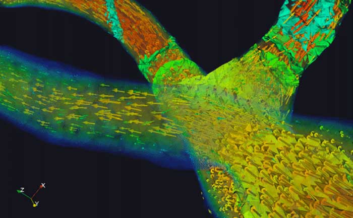
Medical Visualization
Kitware is a leader in scientific visualization, including medical data visualization. Kitware began with the open-source release of the Visualization Toolkit (VTK) in 1996, and that toolkit has become the leading visualization tool in multiple scientific domains, including medical imaging. VTK is capable of generating visualizations of exascale data using supercomputers, heterogeneous data (e.g., genomic as well as image data) using cloud resources, and composite geometric and volume rendered data on desktops; and then VTK can stream any or all of those visualizations to mobile devices, surgical microscopes, and augmented reality / virtual reality systems. Our philosophy is to innovate, promote, and support “pervasive visualization” whereby the data that you need to make a decision is presented to you in an intuitive format, when and where you need it, within your own workflows. Examples of our implementation of pervasive visualizations include the ITK-JupyterWidgets for visualizing data within the Jupyter Lab Python research environment, 3D Slicer for biomedical research data visualization, ParaView Glance for in-browser visualization of a wide variety of scientific data, and ParaView Server for visualization of high-fidelity biomedical simulations of blood flow and/or respiratory air motion.
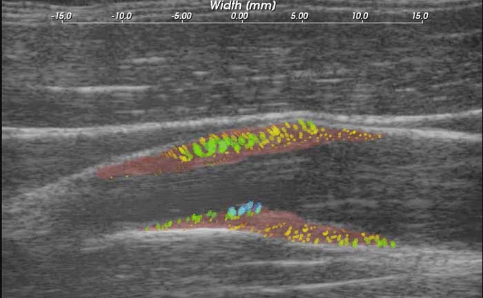
Ultrasound Systems
We are integrating artificial intelligence and deep learning technologies with custom ultrasound and augmented reality hardware to advance the use of ultrasound in a variety of applications. These applications include preclinical and clinical research, pre-hospital patient triage, bedside patient monitoring, and precision needle guidance. Our integrations are enabling less-experienced operators to complete the applications with confidence, in less time, and with expert-level outcomes. These technologies have been transitioned into several consulting projects and commercial products.
3D Slicer-Based Applications
We create custom plugins, SDKs, applications, and software packages using 3D Slicer. 3D Slicer has been used in a variety of medical and basic scientific applications such as dentistry, radiation oncology, surgical planning, and drug development. These custom software applications can be deployed to local hardware or to remote servers using Docker or on tablets. To support reproducible workflows, they can also be integrated with our Girder data management solution or with Jupyter notebooks.
Bioinformatics
Our solutions connect cutting-edge bioinformatics research to a solid, extensible platform, where open science is front and center. We develop data management, analysis management, and visualization applications for a wide range of areas including genomics, metabolomics, and phylogenetics. From browsing and analyzing large histology slides on the web, to stabilizing and sharing new ‘omics algorithms through a robust web application, we have data and analytics covered.
Dental, Craniomaxillofacial, and Musculoskeletal Image Analysis
Our projects and research aim to quantitatively explore how age, disease or treatment affect structures in the skeleton and craniomaxillofacial (CMF) complex. This improved knowledge can help diagnose disease early, plan and measure treatment, and monitor the progression of certain conditions. In particular, we are experts in morphometry analysis, a technique that can be used to quantitatively plan surgery or measure remodeling in the bones. Our musculoskeletal image analysis methods, for example, can quantify bone quality or tooth integrity. We also develop CMF-specific surgical trainers to improve procedural knowledge and surgical proficiency without sacrificing patient safety.
Digital Pathology
We are building a suite of open source web-based informatics tools that manage, visualize, and analyze massive and growing collections of data in digital pathology. The key solutions in the making include Digital Slide Archive (DSA), HistomicsTK, and Large-image. DSA is a web-based platform for the aggregation, management, and dissemination of large collections of whole-slide histopathology images, along with associated clinical and genomic metadata. HistomicsTK serves as both a web-based analytics platform and a standalone Python toolkit. It contains computer vision and machine learning algorithms for the quantitative analysis of whole-slide histopathology images and associated data. Large-image supports the web-based visualization and annotation of large multi-resolution whole-slide histopathology images. It also includes a Python API for reading/writing these images in a tiled fashion.
Image Guided Intervention and Surgical Planning
We develop image-guided intervention and surgical planning applications that replace traditional surgery and invasive procedures with minimally invasive techniques that incorporate medical imaging to guide the intervention. Patients prefer these procedures to open surgeries because they are typically less traumatic to the body and result in faster recovery times. Technological advancements in medical imaging, registration algorithms, visualization technologies, and tracking systems are driving forces behind increased adoption of these procedures by physicians. Software is an integral part of these image-guided intervention systems. Whether it is for interfacing with a tracking device to collect position information from surgical instruments, integrating intraoperative and pre operative images, or generating a 3D visualization to provide visual feedback to the clinician, software has a critical role. The software platforms we are developing at Kitware are playing a major role in increasing the pace of research and discovery in image-guided intervention systems by promoting collaborations between clinicians, biomedical engineers, and software developers across the globe.
Medical Image Analysis
Our expertise in the development of custom image analysis algorithms spans brain morphology assessment associated with mental disorders, tumor volume estimation for clinical trials, vessel modeling for stroke and tumor microenvironment research, multiparametric MRI prostate cancer assessment, deep learning for interpreting histology images, and a number of other applications. Building on our role in the creation and maintenance of libraries such as the Insight Toolkit (ITK) and applications such as 3D Slicer, we lead and partner on basic research grants, small business grants, and development contracts for the National Institutes of Health and the Department of Defense. These encompass nearly every aspect of medical image segmentation, registration, quantification, and computer-aided diagnosis. In addition to working on grants and contracts, we can extend ITK and 3D Slicer with new algorithms to speed the deployment of pre-clinical and clinical products, as well as to collaborate on research investigations.
Medical Visualization
Kitware is a leader in scientific visualization, including medical data visualization. Kitware began with the open-source release of the Visualization Toolkit (VTK) in 1996, and that toolkit has become the leading visualization tool in multiple scientific domains, including medical imaging. VTK is capable of generating visualizations of exascale data using supercomputers, heterogeneous data (e.g., genomic as well as image data) using cloud resources, and composite geometric and volume rendered data on desktops; and then VTK can stream any or all of those visualizations to mobile devices, surgical microscopes, and augmented reality / virtual reality systems. Our philosophy is to innovate, promote, and support “pervasive visualization” whereby the data that you need to make a decision is presented to you in an intuitive format, when and where you need it, within your own workflows. Examples of our implementation of pervasive visualizations include the ITK-JupyterWidgets for visualizing data within the Jupyter Lab Python research environment, 3D Slicer for biomedical research data visualization, ParaView Glance for in-browser visualization of a wide variety of scientific data, and ParaView Server for visualization of high-fidelity biomedical simulations of blood flow and/or respiratory air motion.
Ultrasound Systems
We are integrating artificial intelligence and deep learning technologies with custom ultrasound and augmented reality hardware to advance the use of ultrasound in a variety of applications. These applications include preclinical and clinical research, pre-hospital patient triage, bedside patient monitoring, and precision needle guidance. Our integrations are enabling less-experienced operators to complete the applications with confidence, in less time, and with expert-level outcomes. These technologies have been transitioned into several consulting projects and commercial products.
Did we miss you at the booth?
Here are some helpful resources.
-
PDF
Glance Handout
Visualize your data on the Web
-
PDF
3D Slicer Handout
Powerful tools for medical image informatics, processing, and 3D visualization
-
PDF
VTK Handout
The premiere visualization system
-
PDF
ITK Handout
Extensive suite of tools for image analysis
Working with Kitware
Kitware can work closely with you and your team to customize our platforms. We aim to deliver innovative solutions that not only meet your requirements, but connect with your existing tools and integrate into your workflows. Contact us to learn more about working with Kitware.