BANFF-AID: AI-powered Banff Scoring

Before donated organs can be used, pathologists assess histologic features (including several features covered by Banff lesion scores) based on their overall health and whether key chronic injury indicators are discovered. Unfortunately, this evaluation is largely subjective, resulting in conservative decision-making and the discarding of viable donor organs. The scoring process is also very time-consuming and monotonous, leading to increased errors and worker burnout.
To address these challenges, Kitware partnered with Icahn School of Medicine at Mount Sinai, Emory University, University of Pittsburgh, and University of Alberta on a Phase I SBIR called BANFF-AID: Banff Automated Nephrology Feature Framework – Artificial Intelligence Diagnosis. BANFF-AID is a first-of-its-kind, AI-powered software that automates Banff Lesion Scoring. This software saves time, reduces human error, and improves precision and clinical outcomes.
BANFF-AID was developed as a web-based workflow developed specifically for kidney biopsies. It delivers continuous (non-discretized) scoring of histologic features by integrating custom AI models that perform image segmentation of functional tissue (glomeruli, tubules, arteries, etc.). Our approach ensures that the results are explainable and the method is transparent so pathologists can easily understand how the algorithm computed the lesion score. This approach also empowers pathologists to correct the feature segmentation if it was inaccurate.
During Phase I, we focused on analyzing frozen sections, which represent the standard of care in histopathology for transplant biopsies. Using frozen sections is faster (~20 minutes) than standard formalin-fixed paraffin-embedded (FFPE) sections (multiple hours). With BANFF-AID, we computed scores for arteriosclerosis, glomerulosclerosis, and IFTA (interstitial fibrosis and tubular atrophy) from H&E (hematoxylin and eosin) stained tissue with more precise definitions than the traditional process.
Developing BANFF-AID: The Technical Details
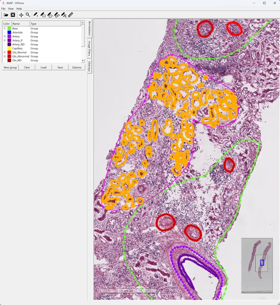
In Phase I, we used 100 annotated H&E images to develop the model. HistomicsTK, Kitware’s open source platform for histopathology, served as the basis for this work. We adapted the University of Florida Multi-Compartment-Segmentation model based on detectron2, which provided a significant development advantage over starting from scratch. Since their model was trained on PAS-stained FFPE images, we retrained it on the same 100 frozen H&E images. Our training started from their final checkpoint. Initial training runs confirmed this approach outperformed starting from standard pre-trained weights. Pre-trained weights allow transfer learning from a large number of natural images contained in the COCO dataset.
Our data processing addressed complex format conversion requirements and partial-annotation challenges. Our work with ASAP XML and Aperio XML formats for WSI segmentations identified specific areas for improvement, particularly tubule detection, where only a small fraction of training images contained tubule annotations. These findings provide clear direction for Task 1 improvements in Phase II.
Building on these insights, we successfully delivered a functional Banff score computation system with dual processing capabilities for individual interactive analysis and high-throughput batch processing. All available frozen H&E images (Mount Sinai + KPMP) were processed and deployed on AWS HistomicsUI for pathologist review and validation.
Evaluating the Results of BANFF-AID
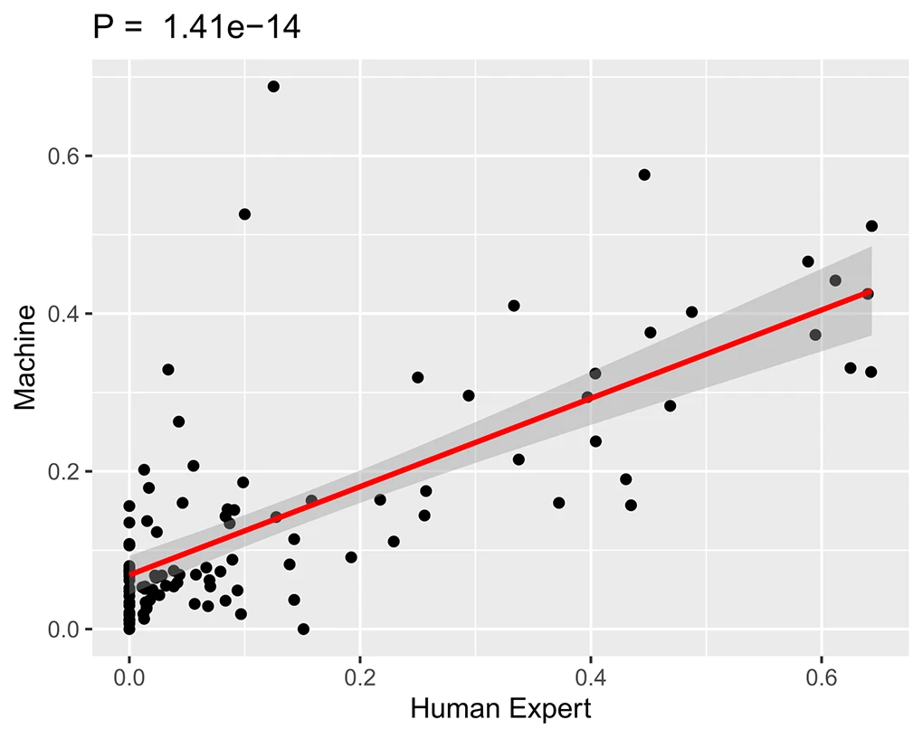
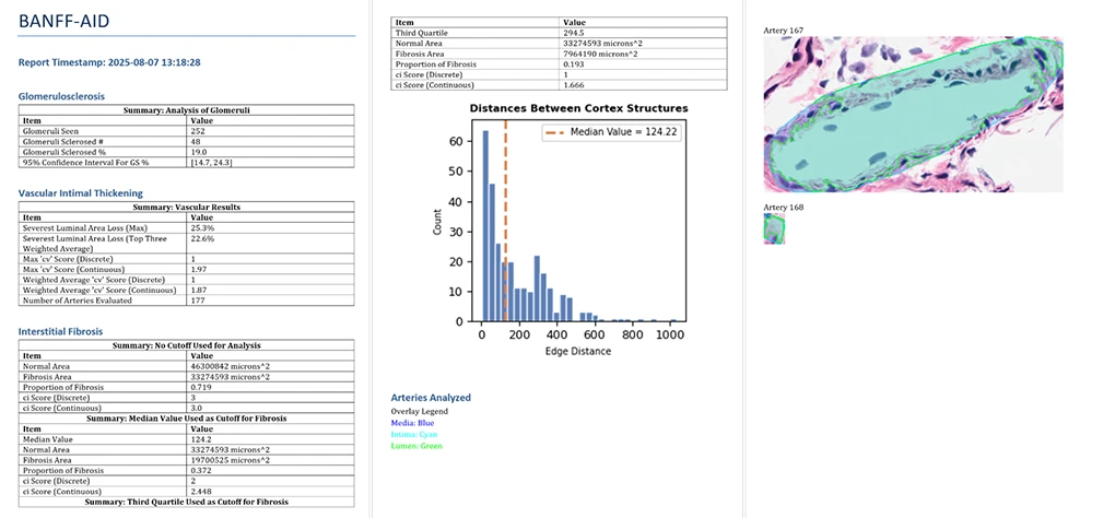
As part of this initial project, we also collaborated with pathologists to conduct qualitative and quantitative validation of BANFF-AID. Figure 2 presents a quantitative correlation analysis of glomerulosclerosis, while Figure 3 shows sample report pages illustrating our current output format. Pathologist feedback provided valuable guidance for Phase II enhancements, including:
- Improving segmentation precision.
- Restructuring reports to highlight critical organ health metrics on the first page with detailed analyses on subsequent pages.
- Refining the terminology to better reflect diagnostic confidence levels (e.g., “interstitial fibrosis” to “interstitial expansion”).
The Future of BANFF-AID
Future phases of development will expand and further optimize the BANFF-AID technology for commercial deployment, focusing on pre-transplant donor kidney assessment with retrospective clinical validation. We intend to add additional collaborations with clinical testing centers and partner with the University of Florida and the University of Nebraska Medical Center.
For Phase II, our comprehensive approach will address two critical needs: reducing viable donor kidney waste by 30% and saving pathologists 30-40% of review time.
Once fully developed, we are confident that this technology would be instrumental for high-volume transplant centers, organ procurement organizations, reference laboratories, research facilities, and pharmaceutical companies developing transplant therapeutics.
Want to learn more?
If you are interested in learning more about this technology, please contact our team.
Acknowledgements
Research reported in this publication was supported by the National Institute Of Diabetes And Digestive And Kidney Diseases of the National Institutes of Health under Award Number R43DK141305. The content is solely the responsibility of the authors and does not necessarily represent the official views of the National Institutes of Health.