SPIE Medical Imaging Papers Accepted

Congratulations to our collaborators and co-workers for three papers accepted to the 2013 SPIE Medical Imaging Conference!
Patient-specific port placement for laparoscopic surgery using atlas-based registration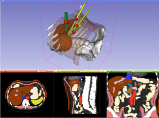
Andinet Enquobahrie, Vikas Shivaprabhu, Stephen Aylward, Julien Finet, Kitware, Inc. (United States); Kevin Cleary, Children’s National Medical Ctr. (United States); Ron Alterovitz, The Univ. of North Carolina at Chapel Hill (United States)
Abstract: Laparoscopic surgery is a minimally invasive surgical approach, in which abdominal surgical procedures are performed through trocars via small incisions. Patients benefit by reduced post-operative pain, shortened hospital stays, improved cosmetic results, and faster recovery times. Optimal port placement can improve surgeon dexterity and avoid the need to move the trocars, which would cause unnecessary trauma to the patient. We are building an intuitive open source visualization system to help surgeons identify ports based on image data and extracted models from abdominal CT image. Our method involves intuitive port placement in abdominal atlas image data and transferring the port locations to individual patients avoiding the need for manual port placement in every patient. Tool maneuverability and target reachability can be tested using the visualization system. In a follow up work, we plan to use the transferred ports as starting point for further optimization of the port locations by formulating a cost function that will take into account factors such as tool dexterity and likelihood of collision between instruments
3D of brain shape and volume after cranial vault remodeling surgery for craniosynostosis correction in infants
Beatriz Paniagua, Omri Emodi, Jonathan Hill, The Univ. of North Carolina at Chapel Hill (United States); James Fishbaugh, The Univ. of Utah (United States); Luiz Pimenta, The Univ. of North Carolina at Chapel Hill (United States); Stephen Aylward, Enquobahrie Andinet, Kitware Inc. (United States); Guido Gerig, The Univ. of Utah (United States); John Gilmore, John van Aalst, Martin Styner, The Univ. of North Carolina at Chapel Hill (United States)
Abstract: Craniosynostosis is a birth defect that causes one or more sutures on an infant’s skull to close prematurely. Corrective surgery focuses on returning the skull to a normal shape. Functional problems caused by craniosynostosis can improve after surgical correction, but a post-surgical analysis of brain development in comparison with age-matched healthy controls is necessary to assess surgical outcome. Volumes and shape metrics in single suture craniosynostosis patients were larger than age-matched controls for pre- and post-surgery. The use of 3D shape and volumetric measurements show that brain growth is not normal in patients with single suture craniosynostosis.
Accelerating ultrasound image analysis research through publically available database
Vikas Revanna Shivaprabhu, Andinet Enquobahrie, Zach Mullen, Stephen Aylward, Kitware, Inc. (United States)
Abstract: Ultrasound (US) is widely used intra-operatively to provide real-time feedback in image guided intervention procedures. Registration of pre- and intra-operative images is challenging and significant preprocessing is required because of the poor signal-to-noise ratio of US images. The amount of preprocessing required can be reduced by incorporating US physical imaging process. However, progress in this research is hampered due to lack of publicly available database for training and testing image analysis algorithms that take in to consideration ultrasound physical process. We present here a new database to archive and distribute US images at different image acquisition parameters that contains tracking information of the transducer in addition to the 2D ultrasound image slices.
Congratulations, very nice work!