Building Software for the Next Generation of Bone Imaging
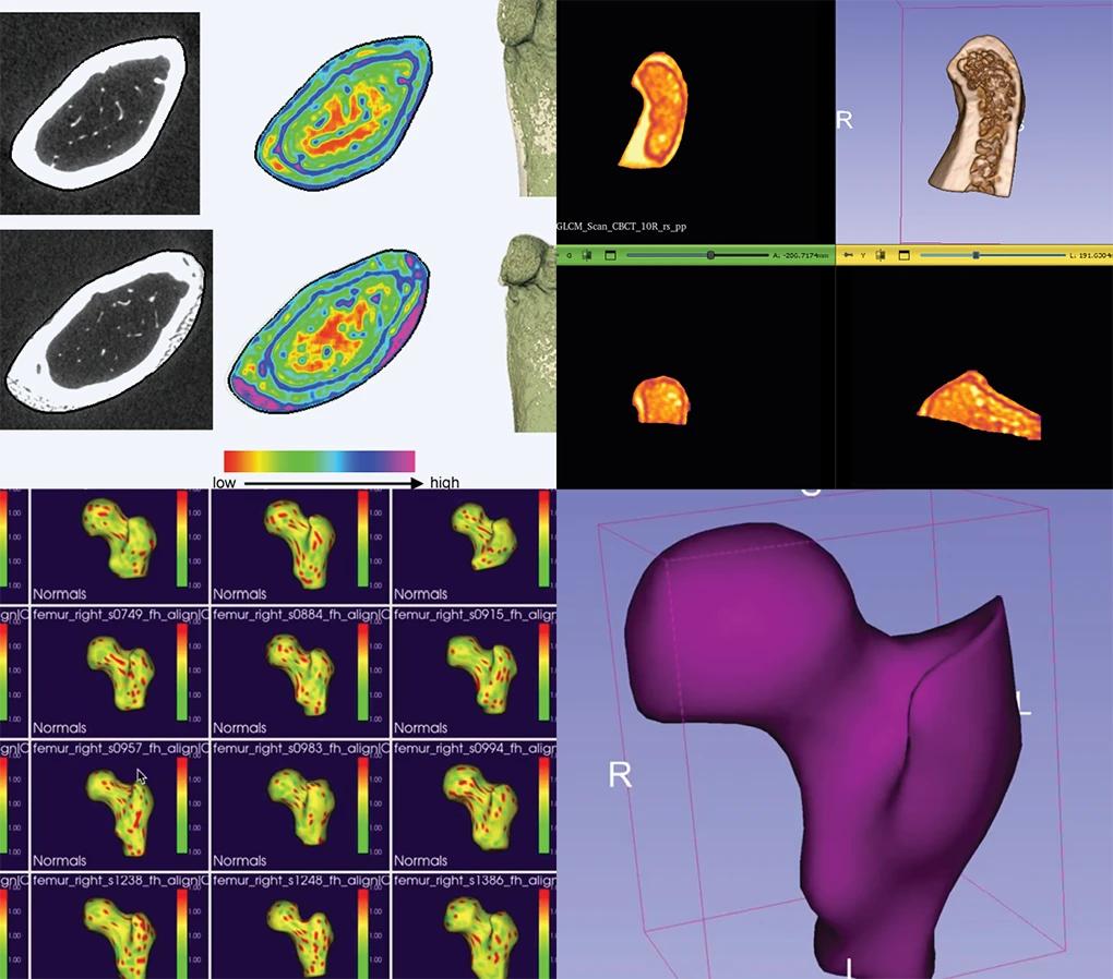
Bone imaging continues to evolve as new imaging modalities, quantitative biomarkers, and AI-assisted analysis expand what researchers can measure and understand. These advances are creating new opportunities across musculoskeletal research. They are also increasing the need for software that can support increasingly complex imaging studies.
Software Challenges in Modern Bone Imaging
Today’s bone imaging landscape is highly diverse. Researchers work with imaging modalities, including microCT, HR-pQCT, WBCT, FUEL CT, high-speed stereoradiography, and MRI. Each modality brings different data formats, analysis methods, and visualization requirements.
Researchers often rely on multiple software applications to complete a single study. Commercial platforms such as Dragonfly, Mimics, VirtualHip, Geomagic, and Analyze each address specific analysis needs. BoneJ provides open source bone morphometry through ImageJ/Fiji. Many research groups also rely on custom MATLAB scripts to perform specialized analyses. Each of these tools addresses a specific need. However, few provide a unified solution across multiple imaging modalities, often requiring researchers to move between several applications to complete a study.
For organizations developing imaging software or supporting large research programs, these disconnected workflows can increase development time and make it more difficult to introduce new analytical capabilities.
Software Requirements Continue to Grow
As research programs expand, software requirements often change. As organizations adopt new imaging modalities and analytical methods, software often needs to:
- Support additional imaging modalities
- Develop custom quantitative analysis algorithms
- Integrate AI-based analysis
- Build browser-based or collaborative applications
- Adapt software to new research questions
Many existing software platforms focus on specific imaging modalities or analysis tasks. As organizations develop new analytical methods or adopt additional imaging modalities, they may need software that can be extended rather than replaced.
Custom Scripts Have Their Limits
Some organizations solve these challenges by developing custom MATLAB scripts. While these scripts can be effective for specific projects, they are rarely designed to support larger research programs. As projects expand, maintaining, validating, and reusing these scripts across studies becomes increasingly difficult.
Many organizations eventually find that adding another software package isn’t enough. They need software that can evolve alongside their research. That may mean extending an existing platform, developing a custom application, or building reusable analysis infrastructure.
How Kitware Helps
Kitware can partner with organizations developing bone imaging software and quantitative analysis tools. We have helped organizations extend existing applications, build custom software, and develop solutions that support evolving research needs by leveraging a collection of open source tools that can help you build standardized, reproducible, and extensible bone analysis infrastructure.
For more than a decade, Kitware has developed open source infrastructure for quantitative bone analysis through NIH-funded research programs that address many of these challenges.
We also partner with organizations to adapt these technologies for new applications, integrate them into existing workflows, and develop custom capabilities that address project-specific requirements.
Our work includes:
- Browser-based tools for multimodal 3D bone characterization and quantitative biomarker analysis, enabling high-throughput reproducible workflows across microCT, HR-pQCT and MRI without proprietary licensing costs.
- Computational algorithms for osteoarthritis assessment and subchondral bone analysis, providing quantitative biomarkers that go beyond the morphological measurements available in commercial platforms.
- The Bone Texture Tool for 3D Slicer, which extracts quantitative texture features from subchondral bone, can replace manual or semi-automated approaches for bone analysis with reproducible quantitative pipelines
- SlicerSALT, which provides statistical shape modeling for longitudinal analysis and large-scale morphometric studies, enables population-level bone geometry analysis that custom scripts will not be able to perform at scale.
- HistomicsTK, which supports web-based computational pathology workflows that complement imaging biomarkers with quantitative tissue analysis that can close the loop between macroscopic structural changes and cellular-level disease mechanisms .
These technologies demonstrate the types of software solutions we develop for our partners. Beyond individual tools, a key strength of Kitware’s approach is the ability to combine these building blocks into cohesive, end-to-end workflows tailored to your specific research or clinical requirements.
Because all of these platforms are built on shared open source infrastructure, they are designed to work together. We build solutions that align with your technical requirements and research objectives.
Connect with our Experts
Bone image analysis continues to move toward AI-assisted segmentation, automated biomarker extraction, multimodal data integration, and large longitudinal studies. As these capabilities become more common, organizations will need software that can integrate new algorithms, support larger datasets, and adapt to changing research priorities.
Whether you’re modernizing an existing platform, developing a commercial imaging application, or building new quantitative analysis capabilities, Kitware can help. Contact our team to discuss your project and explore software solutions tailored to your research and product development goals.